Studies on Stem Cells Research and Therapy
Cytokine Production by Circulating Endothelial Progenitor Cells before and after G-CSF Mobilization
Alexander Lykov*, Olga Poveschenko, Natalia Bondarenko, Alexander Poveschenko, Irina Kim, Eugenie Pokushalov, Alexander Romanov and Vladimir Konenkov
Cite this as
Lykov A, Poveschenko O, Bondarenko N, Poveschenko A, Kim I, et al. (2016) Cytokine Production by Circulating Endothelial Progenitor Cells before and after G-CSF Mobilization. Studies on Stem Cells Research and Therapy 2(1): 001-006. DOI: 10.17352/sscrt.000006Objective: Bone marrow-derived circulating endothelial cells (EPCs) may migrate in ischemia zone, to stimulate resident progenitor cells to proliferation, differentiation and migration in a damage zone, and reduce an ischemia zone through formation of new vessels. Granulocyte colony stimulating factor (G-CSF) is well established to mobilize hematopoietic stem cells and might, thereby, also increase the pool of endogenously circulating EPCs. EPCs secrete pro-angiogenic factors. Therefore, we investigated the effects of G-CSF administration on mobilization and functional activities of blood-derived EPC in patients with chronic ischemic heart disease (CIHD).
Methods and Results: Ten patients with CIHD receive 300 μg per day subcutaneous G-CSF injection for 5 days. The number of EPCs, colony-forming capacity, tube formation and cytokine release were analyzed before and after G-CSF therapy. At day 5 of G-CSF treatment, the number of circulating CD34+CD45- and CD34+CD133+ and CD34+KDR+ cells significantly increased in patients with CIHD. Also, G-CSF therapy augmented the colony-forming capacity and tube formation by EPCs. Likewise, G-CSF treatment augmented cytokine production by circulating EPCs. Early EPCs and late EPCs produced a wide range of cytokines, which dependent the culture condition (gelatin-loaded or fibronectin-loaded surface of culture flask) and the days of cultivation (on day 8 or on day 16).
Conclusion: G-CSF treatment effectively mobilizes EPCs, which through paracrine factors production may influence at the resident progenitor cells in ischemic zone of heart to stimulate the repair of myocardium through neoangiogenesis.
Introduction
At present, cellular transplantation of autologous progenitor cells is perspective way of therapy of degenerative processes including of patients with chronic ischemia heart disease (CIHD) [1,2]. Bone marrow progenitor cells are able to migrate in ischemia zone, to stimulate resident progenitor cells to proliferation, differentiation and migration in a damage zone, and reduce an ischemia zone through formation of new vessels [1-5]. Effects of progenitor cells through production of a wide range of paracrine factors released from these cells.
In previous studies, we have shown the clinical efficiency of intramyocardial bone marrow progenitor cells injection in patients with CIHD [6,7].
The primary objective of the present study was to characterize the spectrum of cytokine and growth factor production by granulocyte colony stimulating factor (G-CSF) mobilized bone marrow cells on periphery in patients with CIHD.
Materials and Methods
Ethics statement
Institutional Review Board approval and informed consent for sample collection were obtained before collection of all samples. All the procedures specified below were approved by Ethical Committee of Institute of Clinical and Experimental Lymphology and Institute of Circulating Pathology and were accordance with the Helsinki Declaration.
Patients and study design
Ten patients (7 men and 3 women between 55 and 66 years) with CIHD and no-option for revascularization consecutive patients with chronic myocardial infarction and end-stage chronic heart failure under optimized pharmacological therapy (β-blockers, ACE inhibitors/ATR-blockers, diuretics) were treated with subcutaneous injections of 300 μg per day G-CSF (Grasalva) for 5 days subsequently days. Inclusion criteria comprised: a history of myocardial infarction >12 months before the enrollment and a fixed perfusion defect on Tc-99m tetrofosmin SPECT; the class of clinical symptoms of heart failure; non revascularisable patient who is symptomatic on optimal medical therapy left ventricular ejection fraction <35% as determined by two-dimensional echocardiography were recruited from Novosibirsk Research Institute of Circulation Pathology of EN Meshalkin MH RF. The following exclusion criteria were applied: eligibility for per cutaneous coronary intervention, coronary artery bypass grafting, previous valve surgery, surgical remodeling of the left ventricle or cardiac resynchronization therapy; hemorrhagic symptoms, severe renal and liver dysfunction, and the history of malignancy.
Isolation, cultivation, and characterization of endothelial cells
On day 0 and day 5 of the therapy, mononuclear cells (MNC) were isolated from venous blood via Ficoll density gradient centrifugation and 106 cells/cm2 were plated on T75 flasks in Dulbecco modified Eagle medium (DMEM, Gibco) with 3mM glutamine, 80 μg/mL gentamycin, Hepes buffer and 10% of fetal calf serum (FCS). After 3 days of culture, non-adherent cells were removed by washing with phosphate buffered solution (PBS) and adherent cells underwent for cultivation on 0.2% gelatin-coated or 0.01% fibronectin-coated flasks during next 16 days. Cultures were maintained by media exchange every 3-4 day. The appearance of well-circumscribed colonies with a cobble-stone morphology was monitored daily. Identification of early and late EPCs was characterized by the morphology and time of culture (early EPCs appearance on day 8 and late EPCs appearance on day 16).
Human umbilical vein endothelial cells (HUVECs) were isolated from freshly harvested umbilical cords. Briefly, the vein was flushed with sterile PBS and then incubated with 0.1% collagenase type I (Sigma-Aldrich) for 20 minutes at 37 °C. The digestion product and subsequent PBS wash were collected and centrifuged. The cell pellet was resuspended in M-199 (Invitrogen) supplemented with 10% FCS plated onto T25 flasks, and allowed to attach overnight. PBS was used to wash away any red blood cells the following day. Media was changed every 2–3 days. EA.Hy926 cells, a human endothelial cell line, were a kind gift from Dr. Cora-Jean S. Edgell (Department of Pathology, University of North Carolina, USA). Cells were cultured in high-glucose DMEM with 10% FCS. The cultures were maintained at 37 °C in a humidified atmosphere (5% CO2 and 95% air), and the medium was renewed every 2-3 days until confluence.
Conditioned medium preparation
To produce human PB-MNC conditioned medium, cells were cultured for 48 hours in growth medium DMEM with 10% FCS. To produce human EPCs conditioned medium, cells were cultured for 8 day and for 16 day in gelatin-coated or fibronectin-coated tissue flask in growth medium DMEM with 10% FCS. To produce HUVECs and EA.hy 926 conditioned medium, cells were cultured for 72 hours in growth medium DMEM with 10% FCS. The conditioned medium was collected and centrifuged to harvest a cell-free solution.
Colony assay
After 3 days of culture, adherent cells were washed with PBS and detached with EDTA; 5x10⁴ isolated EPCs were seeded in methylcellulose plates (Methocult GF H4434) with 100 ng/mL human recombinant VEGF. Plates were studied under phase contrast microscopy, and colonies were counted after 14 days of incubation. Colonies that contained 50 and more cells were defined as endothelial cell colony forming unit (EC-CFU).
Tube formation assay
The formation of vascular-like structures by PB-MNC was assessed on growth factor reduced Matrigel (Becton Dickenson). PB-MNC were seeded on Matrigel-coated 96-well plates at 2x10⁴cells/well in 100 mL of DMEM medium containing 10% FCS and incubated at 37 °C in 5% CO2 for 18 hours. Capillary-like structure formation images were observed using an inverted contrast microscope (Zeiss).
ELISA
To determine cytokine production PB-MNCs (106 cells/mL) were cultured with 10 mg/mL of Con A (Sigma-Aldrich) or 5 mg/mL of LPS (Sigma-Aldrich) or 300 μg/mL of G-CSF (Grasalva) or 33 IU/mL of Epo (Recormon) in 1 mL of growth DMEM medium with 10% FCS in 24-well, flat-bottomed plates at 37 °C in 5% CO2 in duplicate. Cell-free supernatants were harvested after 48 hours of culture and were stored at -70 °C until use. Also, supernatants from EPCs, HUVECs and EA.hy 926 cells were collected to establish cytokine production. ELISA kits (TNF-α, IL-10, IL-18, IL-8, Epo, G-CSF, VEGF; Vector-Best) were used to measure cytokine levels according to the manufacturer’s protocols.
Flow cytometric analysis
On day 0 and day 5 a volume of 100 μL peripheral blood was incubated for 15 minutes in dark with monoclonal antibodies against human CD34 (Becton Dickenson), followed by phycoerythrin (PE) conjugated secondary antibody, with the fluorescein isothiocyanate (FITC) labeled monoclonal antibodies against human CD45, with the PE conjugated monoclonal antibody against human CD133 (marker of immature endothelial cells - early EPCs) and an antibody against the KDR (marker of mature endothelial cells - late EPCs). After incubation, cells were lysed, washed with PBS, and fixed in 2% paraformaldehyde before analysis.
Nitrite production
Nitrite was measured as an indicator of NO production in supernatants from PB-MNC, EPCs, HUVECs and EA.hy 926. A 100 μL aliquot of the culture supernatant was input in a 96-well plate, and an equal amount of Griess reagent (1% sulfanilamide and 0.1% N-1-(naphthyl) ethylenediamine dihydrochloride in 2.5% H3PO4) was added. The plate was then incubated for 5 min, and the absorbance was measured at 540 nm. The amount of NO was calculated using a sodium nitrite standard curve.
Statistical analysis
The Kolmogorov-Smirnov test was used to test for normal distribution of all variables. Continuous variables are presented as mean ± standard error, if not stated otherwise. All comparisons between groups were performed with the nonparametric Mann-Whitney U test. Statistical significance was assumed if p was < 0.05. The correlation between quantity of circulating EPC and level of spontaneous production of cytokines was measured by Spearman’s rank order correlation coefficient (r). Statistical analysis was performed using Statistica (version 6.0).
Results
Characterization of EPCs
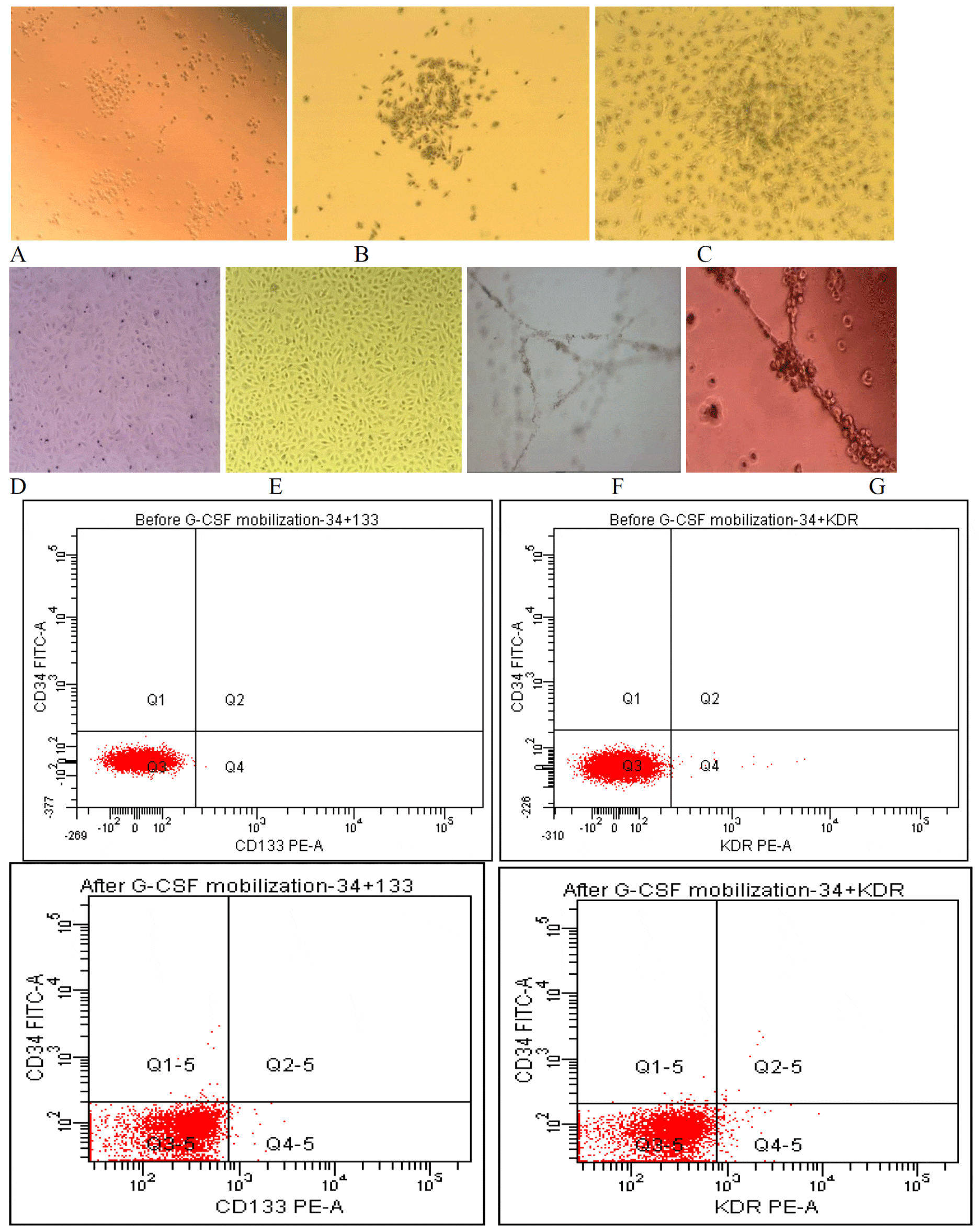
To evaluate the efficiency of G-CSF for mobilization of hematopoietic stem cells in patients with CIHD, 10 patients with CIHD and no-option for revascularization consecutive patients with chronic myocardial infarction and end-stage chronic heart failure and optimized pharmacological therapy received G-CSF doses 300 μg for 5 subsequent days. To evaluate the mobilization EPCs, flow cytometry analysis of the patient peripheral blood was performed for the EPC markers CD34, CD45, and the early marker CD133 and the late marker KDR (Figure 1H). After G-CSF treatment, the number of circulating CD34+ CD133+ cells (phenotype of early EPCs), the number of circulating CD34+ KDR+ cells (phenotype of late EPCs) and circulating CD34+ CD45- cells increased significantly by 2475% (0.004 ± 0.0001% vs. 0.099 ± 0.003%; p < 0.05), by 291% (0.048 ± 0.003% vs. 0.14 ± 0.016%; p < 0.05) and 632% (0.098 ± 0.02% vs. 0.62 ± 0.155; p < 0.05), respectively. To determine the specific mobilization of EPCs by G-CSF, EPCs were cultivated from MNC isolated on day 5 of G-CSF administration. Adherent EPCs were characterized by EC-CFU in a methylcellulose assay. The mobilized MNC that initially seeded were round (Figure 1A). The colonies of early EPCs appeared with the round cells in the centers and typical spindle cells at the periphery after seven days (Figure 1B). At the 18 days, cells (late EPCs) showed characteristic homogeneity and cobblestone-like morphology (Figure 1C) similar to HUVEC (Figure 1D) and EA.hy 926 (Figure 1E). Moreover, circulating EPCs seeded on Matrigel formed capillary-like structures (Figure 1F) as those for EA.hy 926 (Figure 1G).
Cytokine profile
Cytokine profiling revealed that various cytokines were (i.e., TNF-α, IL-10, IL-18, IL-8, Epo, G-CSF, VEGF) secreted by PB-MNC on day 0 and day 5 of therapy (Table 1). Among the cytokines measured, greatest increase in levels of TNF-α, IL-10, IL-18, IL-8, Epo, G-CSF, and VEGF were observed when PB-MNC on day 0 of therapy were co-cultured together in vitro with some stimuli (i.e., ConA, LPS, G-CSF and Epo). On day 5 of therapy PB-MNC when co-cultured in vitro with some stimuli (i.e., ConA, LPS, G-CSF and Epo) greatest increase in levels of TNF-α, IL-10, IL-18, and G-CSF were observed. When levels of cytokines and growth factors produced by PB-MNC on day 0 and day 5 of therapy were compared we observed decrease spontaneous levels production of TNF-α, increase spontaneous levels production of IL-18 and Epo by PB-MNC on day 5 of therapy compare to levels of spontaneous production on day 0 of therapy in patients with CIHD. This indicated that PB-MNC may produce predominantly high levels of important cytokines and growth factors such as TNF-α, IL-10, IL-18, IL-8, Epo, G-CSF, and VEGF. In endothelial cells, eNOS is a constitutively produced protein, required for NO production. To assess whether eNOS induced in PB-MNC by G-CSF represented functional enzyme, cells obtained on day 0 and day 5 of therapy in patients with CIHD were monitored for NO release using nitrite levels as an index of NO production. G-CSF don’t activate of NO production by PB-MNC on day 5 of therapy (Table 1).
We observed positive correlation between quantities of CD34+CD133+ cells in patients with CIHD obtained on day 0 therapy by G-CSF and spontaneous level of IL-18, and VEGF production by mononuclear cells (r= 0.94; p= 0.00004). Also, was established positive correlation between of quantities of CD34+KDR+ cells in patients with CIHD on day 5 of G-CSF therapy and spontaneous level Epo production by mononuclear cells (r= 0.93; p= 0.00009). The negative correlation between quantities of CD34+KDR+ cells in patients with CIHD and spontaneous level of IL-8 production by mononuclear cells (r= -0.85; p= 0.002). Between quantities of CD34+KDR+ cells in patients with CIHD on day 5 therapy by G-CSF and spontaneous level of G-CSF production by mononuclear cells was obtained positive correlation (r= 0.85; p= 0.002). The quantities of circulating CD34+CD133+ cells on day 5 of therapy by G-CSF in patients with CIHD positively correlate with level of NO production by mononuclear cells (r= 0.93; p= 0.00006). These data suggested that the production of some cytokines may be, at least in part, due to the increased quantities of circulating EPC in patients with CIHD.
Cytokine secretion by early or late EPCs
EPCs present in peripheral blood can be at different stages of endothelial differentiation, consequently from PB-MNC can be derived early and late EPCs. Early EPCs occurs during the 2nd weeks of cultivation, and late EPCs appear between 2nd and 3rd weeks of cultivation in vitro. To evaluate the efficiency of gelatin or fibronectin to production by early or late EPCs cytokines and growth factors PB-MNC on day 5 of therapy from patients with CIHD were seeded on 0.2% gelatin-coated and 0.1% fibronectin-coated flasks and cultivated through day 8 (early EPCs) and day 16 (late EPCs). Conditioned medium from early and late EPCs were used to measure the release of different cytokines and growth factors to determine to what extent time of cultivation of PB-MNC the paracrine activity of EPCs. Indeed, on day 8 of cultivation early EPCs on gelatin condition secrete a significant level of IL-8 compare to fibronectin condition (Table 2). In contrary, early EPCs on fibronectin condition secrete a significant level of IL-10, IL-18, Epo and VEGF compare to gelatin condition. Late EPCs on gelatin condition release a significant level of IL-18, Epo and VEGF compare to fibronectin condition. On the other side, late EPCs on fibronectin condition produced a significant level of IL-8 compare to gelatin condition. No significant changes in NO under gelatin or fibronectin condition were observed in EPCs.
Cytokine secretion by EA.hy 926 and HUVEC
Cytokine profiling revealed that various cytokines were secreted by HUVEC and EA.hy 926. Among the cytokines measured, decreased levels of TNF-α, IL-8, Epo and NO produced by EA.hy 926 compare to HUVEC (Table 3). No significant changes in IL-10, IL-18, G-CSF and VEGF production were observed in EA.hy 926 and HUVEC.
Discussion
Ischemic heart disease remains a leading cause of mortality and morbidity [8]. There is a growing interest for cellular therapy, especially bone marrow-derived endothelial progenitor cells, in patients with CIHD, because EPCs not only marker of heart failure, but also contribute to heart repair [2,4-7]. One of the methods of obtained EPCs from bone marrow without aspiration is G-CSF administration which leads to mobilization of progenitor cells from bone marrow [9]. Previous study showed that EPCs secreted a number of cytokines that could stimulate proliferation, migration, and survival of endothelial cells [10-14]. Endothelial progenitor cells promote vasculogenesis and/or ameliorate the process of angiogenesis, thereby improving both regeneration and function of ischemic organs such as the infracted heart [15]. The present study describes the effects of G-CSF on the mobilization and functions including secretory capacity of circulating progenitor cells. The investigated patients with CIHD were treated with a state-of-the-art pharmacotherapy including β-blockers, ACE-inhibitors and diuretics. First of all, we will be sure that G-CSF effectively mobilized progenitor cells in circulation. For this reason, we investigated phenotype of EPCs on day 0 and day 5 during G-CSF treatments. According with finding from other groups, we obtained that G-CSF leads to a mobilization of CD34, CD133 or KDR progenitor cells from the bone marrow in CIHD patients [9]. We demonstrated that the endothelial progenitor cells colony-forming capacity and tube formation increased after G-CSF administration, indicating a potential use this EPCs in cell-based therapy for cardiac repair. Bone marrow-derived EPCs contribute to regeneration of infracted myocardium by enhancement of neovascularization and putative paracrine effects such as secretion of pro-angiogenic factors [7-8,10,14-16]. Our next goal was to investigate whether G-CSF leads to an alteration of the cytokine secretion of EPCs. According with finding from other groups, we obtained that G-CSF leads to decrease of TNF-α, and increase IL-18 and Epo production by PB-MNC in patients with CIHD. It’s known that TNF-α can altered endothelial cells functions [17-19]. Epo have protective effects not only for hematopoietic cells, but also to endothelial cells [16,20,21]. Moreover, Epo can also stimulate proliferation and angiogenesis of endothelial cells that express Epo receptors [12,21]. It’s known that to obtaining early EPCs or late EPCs used fibronectin-coating or collagen-coating [22]. Was showed that during cultivation EPCs with fibronectin up-regulated gene with pro-angiogenic properties (galectin-3) [23]. Therefore, we tested the influence of gelatin or fibronectin condition to cytokine production by EPCs on day 8 and day 16 of cultivation in vitro, which are believed to play an important role for in vivo migration of EPCs to ischemic tissue [3,5,8-11]. We obtained that condition of EPCs cultivation play a significant role on cytokine secretion. So, growing on day 8 EPCs under gelatin condition leads to decrease of IL-10, and IL-18, and Epo, and VEGF production compare to those levels produced by EPCs in fibronectin condition. Whereas, growing on day 16 EPCs under gelatin condition produced a high level of IL-18, and Epo, and VEGF, but reduced the production of IL-8 compare to those cytokine levels secreted by EPC under fibronectin condition. According with finding from other groups, we showed that EPCs secrete a number of cytokines that could stimulate proliferation, migration, and survival of endothelial cells [8,10,14-15]. These results were similar to previous reports [8,10,15]. EPCs secreted a significantly higher level of angiogenic cytokines (IL-8 and VEGF) than did the HUVEC and EA.hy 926. Was estimated that early EPC produced VEGF, G-CSF and IL-8 in significant higher levels than late EPCs. These cytokines can activate adjacent endothelial cells and enhance angiogenesis. In most studies investigated only differences in the levels of cytokine production by early EPCs or late EPCs, but the changes in the levels and the range of cytokines production in the dynamics of maturation of EPCs, and how does the condition of cultivation influences at the range of cytokines production by EPCs wasn’t studied yet. In our study showed that the levels and the range of cytokines production by EPCs depends on the condition of cultivation and time of conditioning in vitro. We believe that this is due to the fact that during mobilization G-CSF of EPCs in the circulation from bone marrow migrated EPCs which are at different stages of maturation and differentiation, and the fact that early EPCs more adherent to fibronectin, while late EPCs stronger adherent to gelatin. On the other hand, with increasing time of cultivation the quantity of late EPCs increased and the quantity of early EPCs reduced, which naturally promotes changes of a range cytokine production. At the present, there is no best approach to identify EPCs. The lack of unique surface markers and heterogeneity of EPCs make it difficult to define EPCs. So, the functional assays as cytokine production and ability to differentiate into endothelial cells should be included for identification of EPCs.
In conclusion, we characterized cytokine production by circulating EPCs in patients with CIHD before and after G-CSF mobilization. The EPCs produced different baseline amounts of cytokine and increased it levels to response to any stimuli (Con A, LPS, Epo, and G-CSF). We obtained differences in cytokine production by early or late EPCs which dependence from culture conditions. We assume that cytokine produced by PB-MNC, also EPCs; by paracrine effects can posse’s survival of injected intramyocardial progenitor cell in patients with CIHD.
- Ichim TE, Solano F, Lara F, Paris E, Ugalde F, et al (2010) Feasibility of combination allogeneic stem cell therapy for spinal cord injury: a case report. Int Arch Med 3: 30.
- Benndorf R, Boger RH, Ergun S, Silva GV, Zheng Y, et al (2011) Imaging Long-Term Fate of Intramyocardially Implanted Mesenchymal Stem Cells in a Porcine Myocardial Infarction Model. PLoS ONE 6: e22949.
- Benndorf R, Böger RH, Ergün S, Steenpass A, Wieland T (2003) Angiotensin II Type 2 Receptor Inhibits Vascular Endothelial Growth Factor–Induced Migration and in vitro Tube Formation of Human Endothelial Cells. Circ Res 93: 438-447 .
- Hristov M, Erl W, Weber PC (2003) Endothelial Progenitor Cells Mobilization, Differentiation, and Homing. Arterioscler Thromb Vasc Biol 23: 185-1189 .
- Hristov M, Zernecke A, Bidzhekov K, Liehn EA, Shagdarsuren E, et al (2007) Importance of CXC chemokine receptor 2 in the homing of human peripheral blood endothelial progenitor cells to sites of arterial injury. Circ Res 100: 590-597 .
- Konenkov VI, Pokushalov EA, Poveschenko OV, Kim II, Romanov AB, et al (2012) Phenotype of peripheral blood cells mobilized by granulocyte colony-stimulating factor in patients with chronic heart failure. Bull Exp Biol Med 153: 124-128 .
- Pokushalov E, Romanov A, Chernyavsky A, Larionov P, Terekhov I, et al (2010) Efficiency of intramyocardial injections of autologous bone marrow mononuclear cells in patients with ischemic heart failure: a randomized study. J Cardiovas Transl Res 3: 160-168.
- Maltais S, Perrault LP, Ly HQ (2010) The bone marrow-cardiac axis: role of endothelial progenitor cells in heart failure. Eur J Cardio-thorac Sur 39: 368-374 .
- Honold J, Lehmann R, Heeschen C, Walter DH, Assmus B, et al (2006) Effects of Granulocyte Colony Stimulating Factor on Functional Activities of Endothelial Progenitor Cells in Patients With Chronic Ischemic Heart Disease. Arterioscler Thromb Vasc Biol 26: 2238-2243 .
- Hur J, Yoon CH, Kim HS, Choi JH, Kang HJ, et al (2004) Characterization of Two Types of Endothelial Progenitor Cells and Their Different Contributions to Neovasculogenesis. Arterioscler Thromb Vasc Biol 24: 288-293 .
- Lai Y, Shen Y, Liu XH, Zhang Y, Zeng Y, et al (2011) Interleukin-8 Induces the Endothelial Cell Migration through the Activation of Phosphoinositide 3-Kinase-Rac1/RhoA Pathway. Int J Biol Sci 7: 782-791 .
- Ribatti D, Presta M, Vacca A, Ria R, Giuliani R, et al (1999). Human Erythropoietin Induces a Pro-Angiogenic Phenotype in Cultured Endothelial Cells and Stimulates Neovascularization In Vivo. Blood 93: 2627-2636 .
- Wang QR, Wang BH, Zhu WB, Huang YH, Li Y, et al (2011) An in vitro Study of Differentiation of Hematopoietic Cells to Endothelial Cells. Bone Marrow Res 2011: 846096 .
- Qiao W, Niu L, Liu Z, Qiao T, Liu C (2010) Endothelial Nitric Oxyde Synthase as A Marker for Human Endothelial Progenitor Cells. Tohoku J Exp Med 221: 19-27 .
- Asahara T, Masuda H, Takahashi T, Kalka C, Pastore C, et al. (1999) Bone marrow origin of endothelial progenitor cells responsible for postnatal vasculogenesis in physiological and pathological neovascularization. Circ Res 85: 221-228 .
- Stein A, Knödler M, Makowski M, Kühnel S, Nekolla S, et al. (2010) Local erythropoietin and endothelial progenitor cells improve regional cardiac function in acute myocardial infarction. BMC Cardiovascular Disorders 10: 43-52 .
- Amtchislavskiy E., Sokolov DI, Selkov SA, Freidlin IS (2005) Proliferative activity of human endothelial cell line EA.Hy 926 and it modulation. Tsitologiya 5: 393-403 .
- Starikova EA, Amtchislavskiy EI, Sokolov DI et al. (2003) Change of surface phenotype of endothelial cells under influence pro-inflammatory and anti-inflammatory cytokines. Medizinskaja Immunologia 1-2: 39-48 .
- Drabarek B, Dymkowska D, Szczepanowska J, Zabłocki K (2012) TNF? Affects energy metabolism and stimulates biogenesis of mitochondria in EA.hy926 endothelial cells. Int J Biochem Cell Biol 44: 1390-1397 .
- Zakharov YuM (2009) Cytoprotective effects of erythropoietin. Clinical nephrology 1: 16-21.
- Beleslin-Cokic BB, Cokic VP, Yu X, Weksler BB, Schechter AN, et al (2004) Erythropoietin and hypoxia stimulate erythropoietin receptor and nitric oxide production by endothelial cells. Blood 104: 2073-2080 .
- Medina RJ, O'Neill CL, Sweeney M, Guduric-Fuchs J, Gardiner TA, et al. (2010) Molecular analysis of endothelial progenitor cell (EPC) subtypes reveals two distinct cell populations with different identities. BMC Med Genomics 3: 18 .
- Ahrens I, Domeij H, Topcic D, Haviv I, Merivirta RM, et al. (2011) Successful in vitro Expansion and Differentiation of Cord Blood Derived CD34+ Cells into Early Endothelial Progenitor Cells Reveals Highly Differential Gene Expression. PLoS ONE 6: e23210 .
Article Alerts
Subscribe to our articles alerts and stay tuned.
 This work is licensed under a Creative Commons Attribution 4.0 International License.


 Save to Mendeley
