Global Journal of Fertility and Research
Postpartum Perigenital Hematoma: A Case Report
1Department of Gynecology and Obstetrics, EHS Nouar Fadela, Oran, Algeria
2EHS Ain Tmouchent, Algeria
Author and article information
Cite this as
Sihem D, Labdali K, Yassine BM. Postpartum Perigenital Hematoma: A Case Report. Glob J Fertil Res. 2025; 10(1): 026-029. Available from: 10.17352/gjfr.000029
Copyright License
© 2025 Sihem D, et al. This is an open-access article distributed under the terms of the Creative Commons Attribution License, which permits unrestricted use, distribution, and reproduction in any medium, provided the original author and source are credited.Traumatic complications of childbirth can include perineal, vulvar, vaginal, and cervical tears, as well as uterine hematomas and ruptures. These complications can have a psychological impact on the mother, including increasing the risk of Posttraumatic Stress Disorder (PTSD).
Our article is about a 23-year-old patient who presented postpartum with a perineal hematoma of more than 10 cm.
The purpose of our presentation is to inform health professionals about this condition in order to improve post-partum care.
Puerperal (or perigenital) hematoma is a rare and potentially serious hemorrhagic complication of postpartum. The current incidence of puerperal hematomas is estimated at 1/1000 [1,2]. It constitutes a formidable complication whose frequency is poorly understood, and the treatment is not codified. Few studies have evaluated the risk factors for puerperal hematomas.
Peritoneal hematomas are a collection of blood located in the cellular tissue of the vulva, vagina, or parametrium, probably linked to a vascular lesion and occurring following a tear or surgical intervention such as an episiotomy during childbirth. Different types of puerperal hematomas must be distinguished.
- Vulvar hematoma: the vulva is vascularized by the arteries of the pudendal, inferior rectal, transverse perineal, and posterior labial branches;
- Vaginal hematoma: the vagina is mainly vascularized by branches of the uterine arteries;
- Vulvovaginal hematoma: this represents the majority of hematomas, limited internally by the vagina, externally by the levator ani muscle and its aponeurosis;
- Pelvic hematoma: the one we are studying in this series is located above the pelvic aponeuroses in the retroperitoneal or intraligamentary region.
Early recognition of hematomas is crucial to avoid serious complications in the mother’s health [3].
The diagnosis of pelvic hematoma is suggested by the association of signs of internal hemorrhage associated with a permanent dull and deep pain on abdominal palpation, in particular during the retroperitoneal extension of the hematoma. The occurrence of hypovolemic shock without hemoperitoneum in the absence of externalized hemorrhage is highly suggestive. The physical signs that can lead to the diagnosis are guarding, a deviated uterus, a curvature above the femoral arch, and sometimes a positive. Vaginal examination reveals a renitent or firm lateral outerine infiltration pushing back the vaginal cul-de-sac(s), or even a vaginal hematoma that has spread. In the highest forms, the shaking of the lumbar fossa is painful and can suggest a urinary pathology. When the pelvic hematoma spreads to the perineum, the clinical signs of vulvovaginal hematomas are associated. The pain is often intense, localized to the vagina and labia majora, with tenesmus if the rectum or anal canal is compressed. Inspection reveals a painful swelling of the labia majora or the lateral wall of the vagina, pushing back the vaginal cavity.
The radiological diagnosis of pelvic hematomas is based on abdominopelvic CT angiography. CT is the imaging of choice in emergencies due to its availability and rapid acquisition, which allows, on the one hand, the visualization of active bleeding (usually called “blush”) and, on the other hand, a rapid diagnosis. The hematoma appears as a hyperdense image whose volume, location in the ischiorectal fossae, retroperitoneal and/or perineal extension, and the possible compressive effect on the urinary tract can be specified. The diagnostic criteria used to define a pelvic hematoma in our study were the presence of a pelvic hematoma on CT or, in the absence of CT, a bundle of suggestive clinical arguments cited above.
The management of large hematomas remains the least consensual subject. Indeed, most authors are in favor of systematic intervention by embolization or surgery in this case [4].
We report a case of a large genital hematoma (> 10 cm) treated conservatively. We discuss the benefits of this management and the conditions under which we recommend this type of management.
Observation
This is a 23-year-old primigravida patient, originally and residing in Ain Tmouchent Algeria, with no particular medical and surgical history, personal and family, ORh positive blood group who presented to the maternity ward for a full-term pregnancy in labor, on general examination; patient conscious and cooperative with a general condition preserved apyretic without infiltration of the lower limbs, on obstetric examination normal uterine height, painful uterine contractions with good relaxation and on vaginal examination the presentation was cephalic well flexed resting on a thin, flexible effaced cervix dilated to 08 cm, the estimated fetal weight in the labor room was between 3900 and 4000 g, the vaginal route was accepted, the patient having given birth vaginally with a wide unilateral episiotomy of a live male child Apgar 9/10 weighing 4300 g. The episiorrhaphy was performed in good condition, and the examination of the pelvis was unremarkable when the patient left the delivery room.
Two days after delivery, the patient returned for perineal pain on the contralateral side of the episiotomy. The patient’s general condition was unchanged, with slight arterial hypotension, skin and mucosal pallor and skin tone, and readmission was indicated in order to investigate the etiology of her anemia.
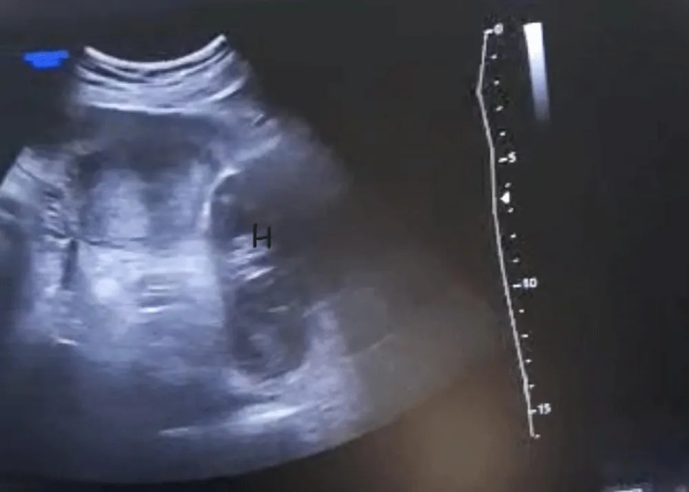
A gynecological examination found, on inspection, a bluish coloration of the contralateral side of the episiotomy, a vulvo-perineal swelling measuring 10 cm in the long axis. The hemodynamic constants were stable with anemia (Hb = 08 g, TP = 100%); the rest of the assessment was without particularity. The ultrasound confirmed the presence of the collection at the level of the pain of significant volume a hypoechoic image (Figure 1), an abdomino-pelvic CT scan was done returning in favor of a pelvic formation of the left pararectal fossa, in intimate contact with the posterior face of the cervix, oval well delimited spontaneously dense without significant enhancement after injection measuring 106 x 74 mm, pushing the rectum to the right.
The patient was transfused with two vials of iso-group iso-rhesus blood under close monitoring of hemodynamic constants, and put on standard treatment: analgesic, anti-inflammatory, and low molecular weight heparin therapy, and uterotonic.
Given the generally stable nature of the patient’s condition and the stability of the size of this hematoma during monitoring, the absence of unbearable pain with improvement in her general condition, we opted for conservative medical treatment (ice pack + NSAID). The evolution was favorable with a progressive reduction in the size of the hematoma until total disappearance at 10 weeks postpartum.
Discussion
This article aims to provide information on the definition, symptoms, diagnosis, and treatment options. The main associated factors were local: presence in all cases of a perineal lesion (episiotomy or tears) and one out of two cases of instrumental extraction (by spatulas). The clinical and radiological signs were recalled [5,6]. Vascular rupture is often venous in the thickness of the vagina with lamination of the wall and diffusion along the fascia [3].
The symptomatology of pelvic hematoma is varied and non-specific. Pain is the earliest and most constant manifestation. Two other suggestive clinical elements emerge that should alert the clinician. First, acute urinary retention, which constitutes an early (in the first hours) and frequent (one third of cases) clinical sign. It is favored by the diffusion of the hematoma in the subperitoneum and the space of Retzius. Second, a clinical picture of hypovolemic shock without hemoperitoneum and without externalized hemorrhage, as well as the discordance between a moderate vulvovaginal hematoma and hemodynamic instability, should also lead to the search for a pelvic hematoma. Different risk factors have been described, such as primiparity, maternal age, birth weight [6], as well as the presence of perineal lesions. Saleem and Rydhstrom recorded 1418 cases of vaginal hematomas from the Swedish national birth register, or 1/1242 births [6]. In our case, we note the importance of the fetal factors: the weight of the child, a feto-pelvic disproportion. Richard et al. report a case of pelvic hematoma after pudendal block whose origin is attributed to a traumatic venous wound of the pudendal pedicle [7].
As with any postpartum hemorrhage, the management of these patients must be multidisciplinary (anaesthetists, obstetricians, and radiologists), including an initial assessment of the severity and confirmation of the cause of the hemorrhage during a pelvic gynecological examination. In the case of a debilitating cervico-vaginal wound not controlled by local treatment or an extensive pelvic hematoma, arterial embolization appears to be the first solution to consider [2,7,8]. It has the advantage of being less invasive than surgery, of localizing the bleeding, and, above all, of preserving the compressive effect of the hematoma left in place, which is not possible with the surgical approach.
After embolization, close monitoring is essential (post-intervention monitoring, resuscitation, or intensive care). In the event of failure of the initial embolization, rapid intervention must be possible (new embolization or surgical hemostasis procedure). Bilateral surgical ligation of the hypogastric arteries remains the reference technique in the second line [7].
The limiting factor in the implementation of this therapeutic strategy is the need to have a 24-hour interventional radiology center. If transfer to a specialized center is considered, certain conditions must be met [6]: medicalized transfer (SMUR type), stable hemodynamic state, and relatively short transfer time. Direct pelvic or retroperitoneal surgical approach should be avoided. However, it seems reasonable to propose the evacuation of a vaginal hematoma associated with the pelvic hematoma when it is greater than 10 cm [5] or when there is significant painful symptomatology [9].
However, in the case of large hematomas (> 8 cm), practices differ. In fact, in this case, most authors systematically recommend active management based on surgery and/or embolization [10]. Other teams, including ours, opt instead for conservative management when the patient is clinically and biologically stable [3,11]. This attitude is argued by the fact that the established hematoma exerts pressure on the surrounding tissues, which can collapse the bleeding vessel and thus allow hemostasis. Evacuating this stable hematoma increases the risk of recurrence of bleeding [12].
Conclusion
Pelvic hematoma is a rare complication of postpartum that deserves to be better understood by practitioners. This non-externalized hemorrhage is often diagnosed late in the face of a severe clinical picture of hypovolemic shock. This is achieved by paying attention to the clinical picture (pain, acute urinary retention, hypovolemic shock without hemoperitoneum, moderate vulvovaginal hematoma with unstable hemodynamics) and identifying certain risk factors common to all puerperal hematomas.
Ethical aspects
The patient’s consent was obtained.
- Parant O, Simon-Toulza C, Capdet J, Fuzier V, Arnaud C, Rème JM. Immediate fetal-maternal morbidity of first instrumental vaginal delivery using Thierry's spatulas. A prospective continuous study of 195 fetal extractions. Gynecol Obstet Fertil. 2009;37(10):780-6. French. Available from: https://doi.org/10.1016/j.gyobfe.2009.07.010
- Boyer L, Dumousset E, Rousseau H. Management of severe post partum hemorrhage with embolization: the French experience. J Radiol. 2006;87(5):549-53. French. Available from: https://doi.org/10.1016/s0221-0363(06)74036-7
- Lédée N, Ville Y, Musset D, Mercier F, Frydman R, Fernandez H. Management in intractable obstetric haemorrhage: an audit study on 61 cases. Eur J Obstet Gynecol Reprod Biol. 2001;94(2):189-96. Available from: https://doi.org/10.1016/s0301-2115(00)00349-3
- Ridgway LE. Puerperal emergency. Vaginal and vulvar hematomas. Obstet Gynecol Clin North Am. 1995;22(2):275-82. Available from: https://pubmed.ncbi.nlm.nih.gov/7651671/
- Zahn CM, Yeomans ER. Postpartum hemorrhage: placenta accreta, uterine inversion, and puerperal hematomas. Clin Obstet Gynecol. 1990;33(3):422-31. Available from: https://doi.org/10.1097/00003081-199009000-00006
- Riethmuller D, Pequegnot-Jeannin C, Rabenja CA, Koeberle P, Schaal JP, Maillet R. A rare cause of postpartum hemorrhage: a genital thrombus. J Gynecol Obstet Biol Reprod (Paris). 1997;26(2):154-8. French. Available from: https://pubmed.ncbi.nlm.nih.gov/9265033/
- Bienstman-Pailleux J, Huissoud C, Dubernard G, Rudigoz RC. Management of puerperal hematomas. J Gynecol Obstet Biol Reprod (Paris). 2009;38(3):203-8. French. Available from: https://doi.org/10.1016/j.jgyn.2008.10.002
- Pelage P, Laissy JP. Management of severe postpartum hemorrhage: indications and techniques of arterial embolization: indications and technique of arterial embolization. J Radiol. 2006;7:533-40. Available from: https://doi.org/10.1016/S0221-0363(06)74034-3
- Villella J, Garry D, Levine G, Glanz S, Figueroa R, Maulik D. Postpartum angiographic embolization for vulvovaginal hematoma. A report of two cases. J Reprod Med. 2001;46(1):65-7. Available from: https://pubmed.ncbi.nlm.nih.gov/11209635/
- Deux JF, Bazot M, Le Blanche AF, Tassart M, Khalil A, Berkane N, et al. Is selective embolization of uterine arteries a safe alternative to hysterectomy in patients with postpartum hemorrhage? AJR Am J Roentgenol. 2001;177(1):145-9. Available from: https://doi.org/10.2214/ajr.177.1.1770145
- Propst AM, Thorp JM Jr. Traumatic vulvar hematomas: conservative versus surgical management. South Med J. 1998;91(2):144-6. Available from: https://doi.org/10.1097/00007611-199802000-00004
- Muthulakshmi B, Francis I, Magos A, Roy M, Watkinson A. Broad ligament haematoma after a normal delivery. J Obstet Gynaecol. 2003;23(6):669-70. Available from: https://doi.org/10.1080/01443610310001607887
Article Alerts
Subscribe to our articles alerts and stay tuned.
 This work is licensed under a Creative Commons Attribution 4.0 International License.


 Save to Mendeley
